
.jpg "LmSmdB")
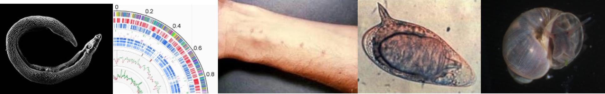
Schistosomiasis, also known as bilharzia, in honour of its discoverer. It is a tropical water-borne disease caused by blood-flukes Schistosomes. It is the second most socioeconomically devastating parastitic disease after malaria in terms of , mortality and morbidity rates. Schistosomiasis, also known as snail fever continues to rank second after malaria in the world's parasitic diseases in terms of prevalence, chronicity, morbidity and mortality rates. Currently, over 210 million individuals in 76 countries have been affected, while close to 800 million people are at risk of contracting this disease. Schistosomes exhibit complex life cycle comprising of morphologically distinct phenotypes in intermediate snail host (molluscs) and definitive human host. Cercariae are the larvae of schistosomes, capable of infecting mammals (human) According to clinical manifestations, two different conditions acute and chronic schistosomiasis are associated with S.mansoni infection. Consequences of chronic infection may range from granuloma formation to fibrosis to severe mal-function of the liver and spleen.
Life cycle of Schistosomiasis
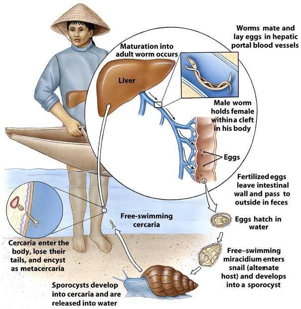
- The eggs of the human-dwelling parasite are emitted in the faeces and into the water, the ripe miracidium hatches out of the egg , in response to temperature, light and dilution of faeces with water.
- The miracidium searches for a suitable freshwater snail (Biomphalaria glabrata, Biomphalaria straminea, Biomphalaria tenagophila or Biomphalaria sudanica) to act as an intermediate host and penetrates it.
- The parasite develops via a so-called mother-sporocyst and daughter-sporocyst generation to the cercaria. The purpose of the growth in the snail is the numerical multiplication of the parasite. From a single miracidium result a few thousand cercaria, every one of which is capable of infecting man.
- The cercaria emerge from the snail during daylight and they propel themselves in water with the aid of their bifurcated tail, actively seeking out their final host. When they recognise human skin, they penetrate it within a very short time.
- This occurs in three stages,
- An initial attachment to the skin.
- Cercaria creeps over the skin searching for a suitable penetration site, often a hair follicle.
- Finally penetration of the skin into the epidermis using cytolitic secretions from the cercarial post-acetabular, then pre-acetabular glands.
- On penetration, the head of the cercaria transforms into an endoparasitic larva, the schistosomule. Each schistosomule spends a few days in the skin and then enters the circulation starting at the dermal lymphatics and venules.
- The schistosomule migrates to the lungs (5-7 days post-penetration) and then moves via circulation through the left side of the heart to the hepatoportal circulation (>15 days) where, if it meets a partner of the opposite sex, it develops into a sexually mature adult and the pair migrate to the mesenteric veins.
- The paired worms move against the flow of blood to their final niche in the mesenteric circulation where they begin egg production (>32 days). The S. mansoni parasites are found predominantly in the small inferior mesenteric blood vessels surrounding the large intestine and caecal region of the host.
- Each female lays approximately 300 eggs a day (one egg every 4.8 minutes), which are deposited on the endothelial lining of the venous capillary walls.
Distribution of Disease
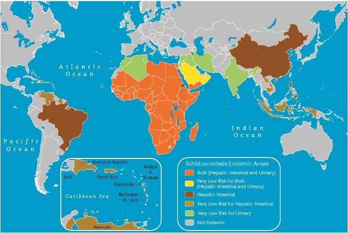
Schistosoma mansoni, one of the prevalent species associated with this disease, is found to occur across much of India, Africa, Middle East and South America.